Advanced cell-based assays for mechanistic and functional characterisation of your assets
Mechanistic and functional assays are essential tools in understanding the underlying biology of diseases and evaluating the efficacy of potential treatments. Our team leverages an extensive range of cellular assay technologies to support our clients at every stage of their drug discovery journey. We can assist with the selection, optimisation and validation of the most promising chemical, biological or cellular leads, to help ensure that you achieve reliable and actionable results. Our expertise and state-of-the-art facilities enable us to deliver tailored solutions that drive your projects forward with precision and confidence.
- Cell proliferation assays: MTT, BrdU incorporation, colony formation, image-based assays (Incucyte, Celligo, HCA, CTG)
- Cytotoxicity assays: LDH, caspase-3/7, annexin V/PI staining
- Migration and invasion assays: wound healing, transwell migration, matrigel invasion
- Signal transduction assays: luciferase or fluorescence-based reporter assays, calcium mobilisation assay
- Apoptosis and cell death assays: TUNEL, mitochondrial membrane potential assay, DNA damage, HCA
- Cell differentiation assays: FACS, alkaline phosphatase staining
- Endocytosis and exocytosis assays: xCelligence-based impedance, fluorescence microscopy, FACS-based cell sorting
- Protein-protein interaction assays: co-immunoprecipitation, FRET, BRET
- Gene expression assays: qPCR, RNA sequencing
- Protein expression: Western blot, Simple Western, in-cell Western, HCA, cell painting, cell morphology, protein localisation, G-Liza, Alpha-Liza, HTRF, IHF, FACS, spatial profiling, global proteomics
- Chromatin immunoprecipitation (ChIP)
- Mass spectrometry-based proteomics
- Proteasomal degradation/activity assays
- Sequencing: preclinical and clinical material, RNA/DNA sequencing, ChIP-seq, ATAC-seq, single-cell RNA-Seq, Perturb-seq
- Metabolic assays: Seahorse XF Analyzer, glucose uptake assay.

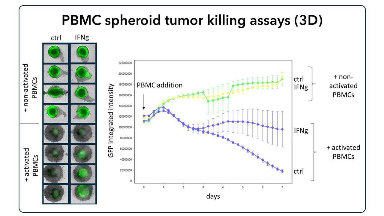
Cancer cells pretreated with IFNgamma are plated on low-attachment plates for spheroid formation. Activated or naïve PBMCs are added to co-culture. Effects of PBMCs on viability of cancer spheroids are detected by image-based readout using an Incucyte.


