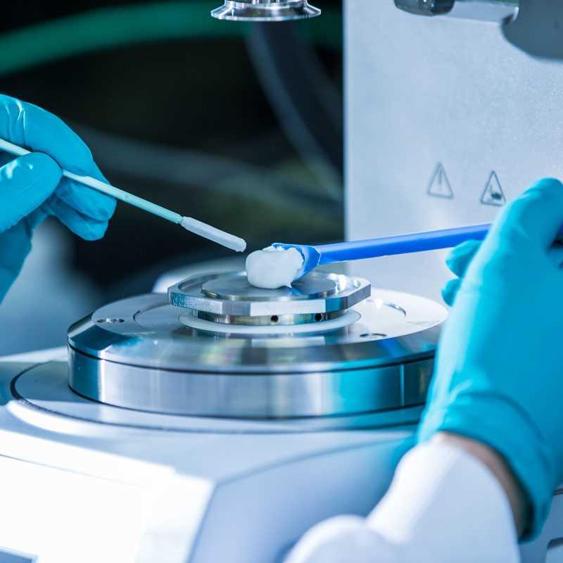
Reducing development risks and increasing the probability of success are key to any project we support. For this reason, we focus on the translational value of models early in the process. Primary cells or tissues from human donors or animals are integral and vital parts of drug discovery and profiling programs.

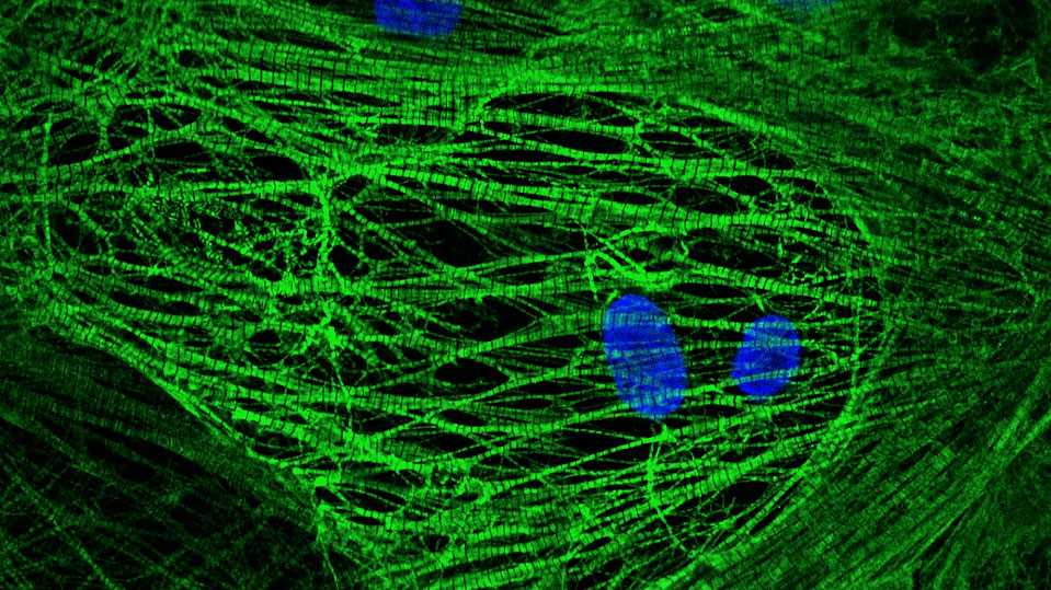
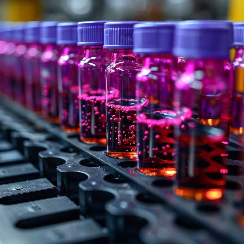
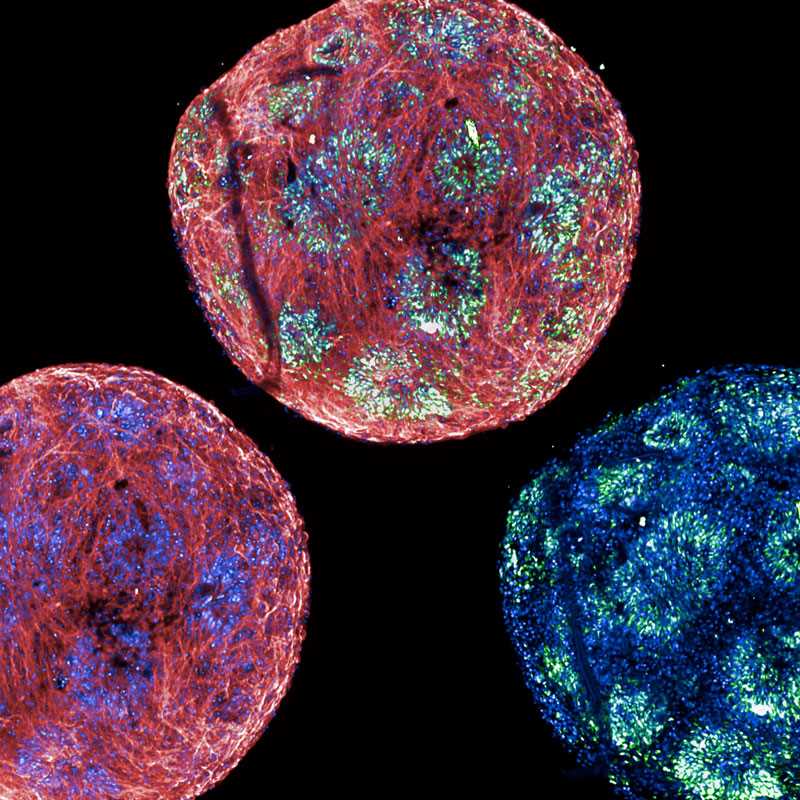
We are constantly expanding our hiPSC-derived cell portfolio in the cardiology and neurology indication spaces.
learn more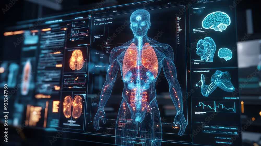
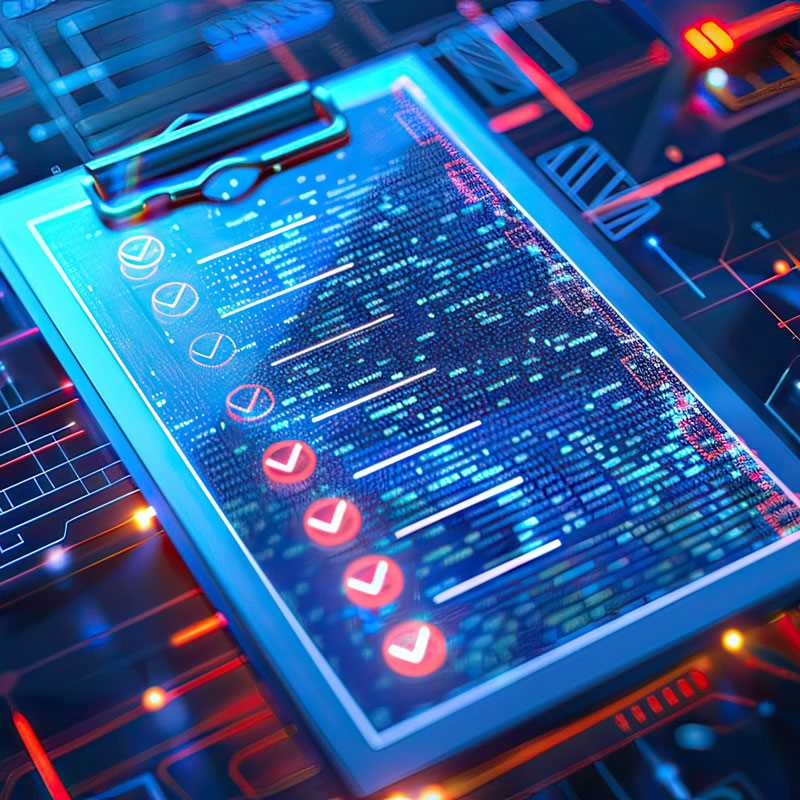
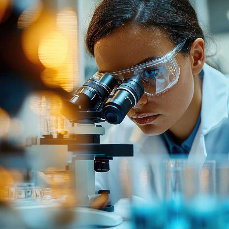
As an AAALAC-certified CRO, we are dedicated to conducting ethical and high-quality in vivo studies.
download