Established hiPSC-derived cell models cardiomyocytes
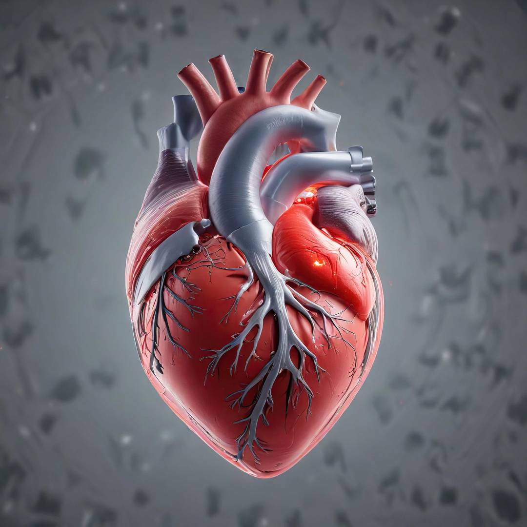
Ventricular cardiomyocytes are the major functional component of heart muscle. We can generate hiPSC-derived ventricular cardiomyocytes that express a range of cardiac and muscle-specific markers that are widely accepted in the field. Each batch of differentiated cardiomyocytes undergoes stringent assessments for purity and viability. We can also perform functional measurements assessing contractility, field potential, signal propagation and local extracellular action potential (LEAP) using the Maestro Pro MEA assay system (Axion BioSystems).
cardiac fibroblasts
Cardiac fibroblasts have an increasingly appreciated role in cardiac homeostasis, with important functions in overall tissue architecture, as well as physiological and pathological remodeling. They are an essential component of our 3D engineered cardiac organoids (ECOs).
cardiac endothelial cells
Endothelial cells are a major component of cardiac vasculature and undergo alterations in response to biochemical and hemodynamic stimuli. They are also directly involved in developmental and pathophysiological responses. Our human iPSC-derived endothelial cells express CD31, CD144 and VEGFR2, form capillary-like structures and retain their ability to form a selective barrier, which is crucial to their function. They are also an optional component of our 3D ECOs.
3D engineered cardiac organoids (ECOs)
It is increasingly accepted that providing cells with the appropriate spatiotemporal context and external cues permits them to advance in their maturation status. We can generate 3D ECOs in multiple geometries (e.g., as spheroids, rings or strips), depending on your desired application. These macro-scale tissues can be tuned in complexity by multiplexing with additional cell types of your choice. By utilising hiPSC-derived ventricular cardiomyocytes and cardiac fibroblasts from the same hiPSC line (donor), we can ensure that we are recapitulating the correct cell–cell interactions unique to that patient. ECOs generate contractile forces and respond to standard reference compounds. We are also able to mimic disease scenarios in vitro, such as localised myocardial infarction with the concomitant release of clinically relevant cell-death markers. We have developed methods that differ from the state of the art to generate ECOs.
nociceptor neurons
Nociceptor neurons are specialised sensory neurons of the peripheral nervous system responsible for detecting noxious stimuli and conveying these as pain signals. They play a significant role in the development of pain-relieving medications and treatments for different pain-related conditions. By targeting nociceptor functions, we provide a model to advance therapies that modulate pain signals at various stages of transmission and processing.
microglia
Microglia are resident immune cells of the central nervous system and play a vital role in maintaining neuronal health and homeostasis. They are increasingly recognised as important targets in neurodegenerative diseases, neuroinflammation and pain-related conditions. Therapeutic approaches aim to regulate microglial activity to suppress harmful overactivation or enhance their neuroprotective functions.
midbrain dopaminergic
Dopaminergic neurons are specialised cells of the central nervous system that produce and release the neurotransmitter dopamine. They play a crucial role in movement and motor control, reward processing, mood regulation and cognition. Dysfunction or loss of dopaminergic neurons can lead to a broad range of severe neurological and psychiatric disorders, including Parkinson’s disease and schizophrenia, making them key targets for the development of therapies for various brain-related conditions.


