Advanced HCA assay systems

Tackling challenging targets

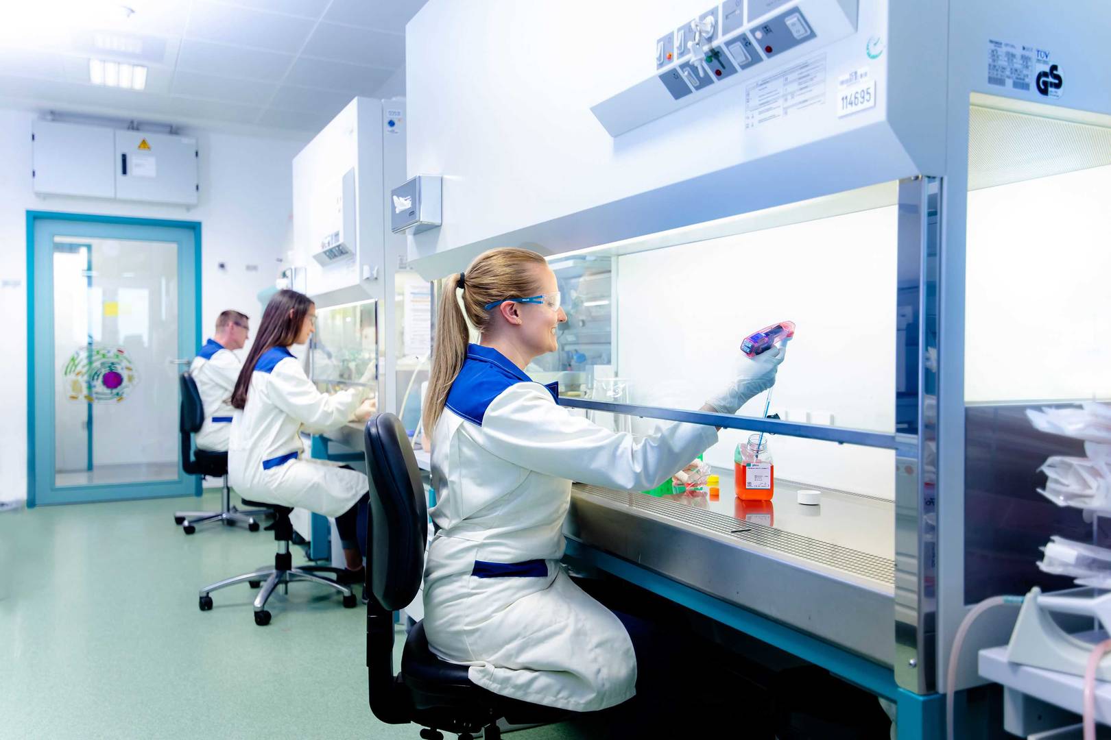
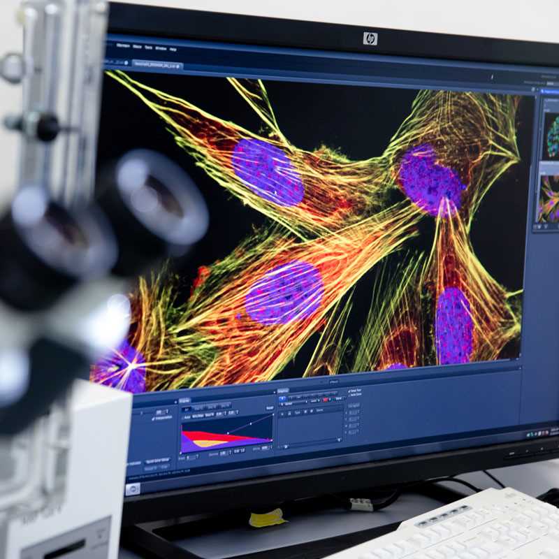
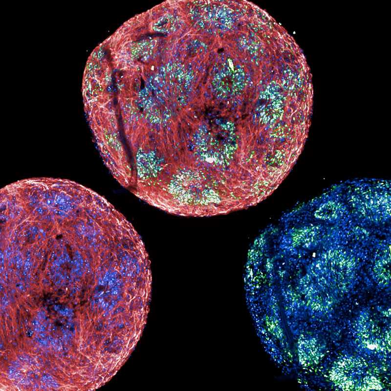
When you are faced with a target that cannot be addressed using traditional approaches, our scientists will support you in implementing advanced HCA assays. These can include complex phenotypic readouts at population or single-cell levels, live-cell kinetics and stem-cell-based systems. Our HCA assays can be multiplexed and made HTS-compatible using small molecules or CRISPR libraries, and can accommodate a wide variety of eukaryotic cells, bacteria and cell-free systems.

A scientist explores imaging data of a high-content assay

Phenotypic readouts

The applicable phenotypic readouts can include, but are not limited to, the sub-cellular (re)localisation of a target, organelles and intra-cellular structures.


Cellular and protein–protein interaction assays

Using fluorescent-based techniques, we develop HCA assays to study protein–protein interactions or interactions between cells. This enables us to identify small molecules that impact these processes. 


Degraders

We identify degrading compounds such as PROTAC®s and molecular glues through high-throughput cellular assays that measure protein stability, utilising immunofluorescence and fluorescent protein tagging to identify novel compounds.


Protein condensates

Together with our clients, we aim to identify protein condensates within cells and discover small-molecule modulators of condensate biology. 


RNA-targeting small molecules

We offer ultra-sensitive visualisation techniques for single mRNA and lncRNA localisation and abundance through fluorescence in situ hybridisation (FISH). Our in-house development of the first dual FISH-HTS ensures precise and reliable results for your research needs. 

Cell painting

At Nuvisan, we offer advanced cell painting services to provide comprehensive cellular phenotyping. We stain various cellular components with fluorescent dyes and capture high-resolution images. The technique labels key structures like the nucleus, mitochondria, cytoskeleton and endoplasmic reticulum with distinct markers. Sophisticated image analysis software extracts detailed morphological features such as shape, texture and spatial organisation. This allows us to identify subtle phenotypic changes due to genetic modifications, chemical treatments or disease states. Ideal for drug discovery and functional genomics, our cell painting services facilitate high-throughput screening and generate rich datasets to help uncover new biological insights and therapeutic targets.


JUMP-Cell painting consortium

Funded by the Bits to Bytes Capital Call of the Massachusetts Life Sciences Center (MLSC), the JUMP-Cell Painting Consortium has developed a data-driven approach to drug discovery using cellular imaging and data analytics. This method addresses a key bottleneck in pharma: determining the mechanism of action, as well as predicting the activity and toxicity of potential therapeutics. The Consortium has created a public dataset to validate and scale this strategy, aiming to make cell images as computable as genomes and transcriptomes. We contributed data to the Consortium and can provide a workflow matching the Consortium's standards for your cell painting needs.

Related topics & resources

High-throughput screening with a plate-based assay

Our HTS solutions

Explore our wide range of screening services and HTS libraries.   

learn more
Screening expert processing a stack of assay plates 

High-throughput HCA

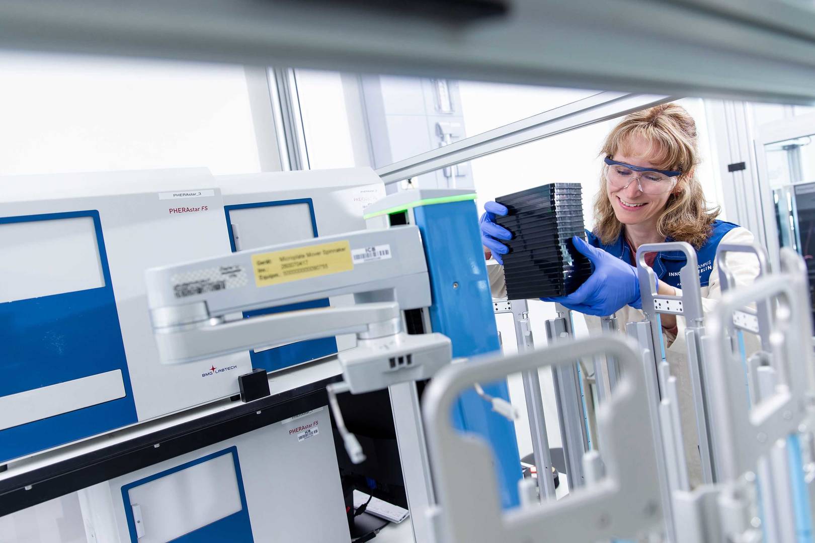
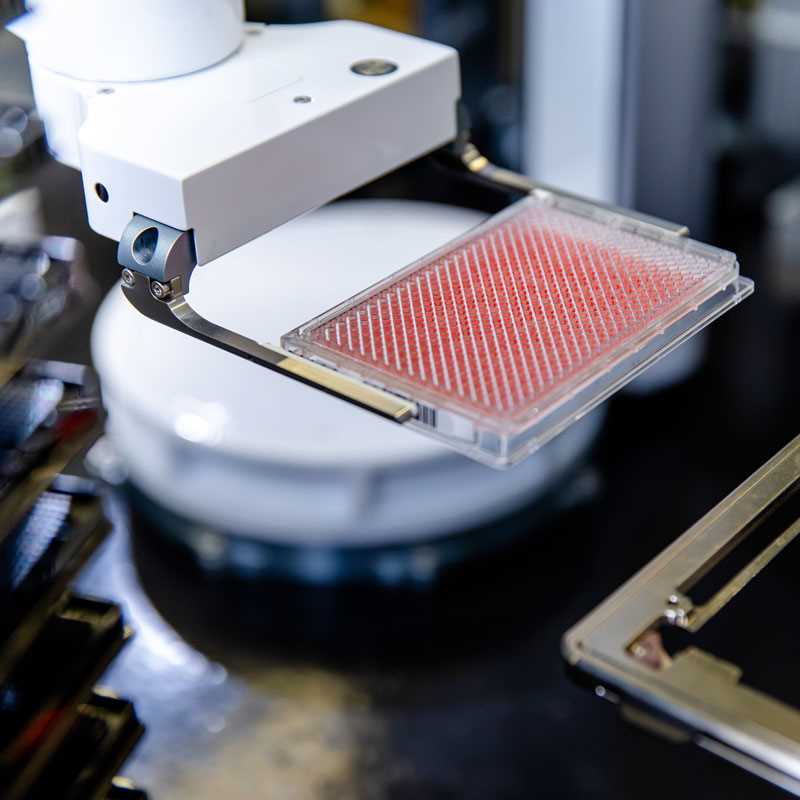
We support our partners and clients with robust automation and miniaturisation of high-content assays. 

learn more
three lab technicians working at the clean bench.

Cell-based assays

Discover our large portfolio of cell-based assays.

learn more